Oral Cyst & Mucocele Removal
for Houston Residents
A complete guide to diagnosing and treating oral cysts and mucoceles. Expert surgical removal at Best Dental in Richmond, TX, serving Houston, Sugar Land, Katy, and Fort Bend County. Fast, comfortable treatment for painful or bothersome oral lesions. Mucocele and fibroma removal is a flat $500 at Best Dental.
On This Page
A complete guide to oral cyst and mucocele diagnosis, treatment, and pricing near Houston, TX.
What Are Oral Cysts & Mucoceles?
Oral cysts and mucoceles are fluid-filled sacs or bumps that develop in the mouth. While they are usually benign, they can cause discomfort, interfere with eating or speaking, and sometimes indicate underlying dental issues that need attention. Understanding what they are and the difference between the two is the first step toward getting the right treatment.
🦴 Oral Cysts
- Develop in jawbone or deep soft tissues
- Often discovered incidentally on X-rays
- Usually painless in early stages
- Grow slowly over months or years
- Can damage surrounding teeth and bone
- Require surgical removal and will not resolve on their own
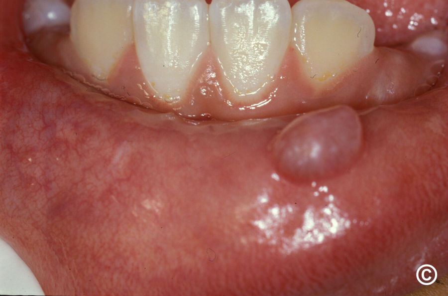
💧 Mucoceles
- Form in soft tissues: lip, cheek, or tongue
- Visible as soft, clear or bluish bumps
- Usually painless unless irritated
- Often appear suddenly after biting lip or cheek
- May rupture and refill repeatedly
- Small ones may resolve; persistent ones need removal
Oral cysts grow slowly and will not go away on their own. Without treatment, they can damage surrounding bone and teeth, making removal more complex over time. Mucoceles form when a salivary gland duct gets blocked or damaged, often from accidentally biting your lip or cheek. Saliva backs up and creates a fluid-filled bubble under the tissue.
At Best Dental in Richmond, TX, our team diagnoses and treats all types of oral cysts and mucoceles in-house. Early evaluation ensures prompt, effective treatment before complications develop.
Any Persistent Bump Deserves an Evaluation
Most oral cysts and mucoceles are completely benign, but any bump or swelling that persists for more than two weeks should be professionally evaluated to confirm the diagnosis and rule out more serious conditions.
Mucocele Removal Cost at Best Dental
Best Dental charges a flat $500 for mucocele and fibroma removal. This covers the complete in-office excision procedure including the removal of the mucocele itself and the associated damaged salivary gland tissue that causes recurrence. There are no separate facility fees or component charges. The $500 fee is the all-in price for a standard mucocele removal at Best Dental in Richmond, TX.
If you have a bump or lesion that needs evaluation, Best Dental charges $89 for a limited exam. This is a focused clinical evaluation of the specific concern, including X-rays as needed, and is not a full new patient exam. The $89 exam fee is separate from and does not apply toward any treatment.
| Procedure | Best Dental Fee | What Is Included |
|---|---|---|
| Mucocele Removal | $500 flat | Complete excision of mucocele and damaged salivary gland tissue. Local anesthesia included. Dissolvable sutures. Post-op instructions. |
| Fibroma Removal | $500 flat | Surgical excision of fibrous tissue growth. Local anesthesia included. Tissue sent to pathology for confirmation. |
| Oral Cyst Removal (small) | Quoted at consultation | Fee depends on cyst size, location, and whether associated teeth require treatment. Transparent quote provided before treatment begins. |
| Oral Cyst Removal (large or complex) | Quoted at consultation | Larger cysts or those requiring bone grafting are quoted individually. Written estimate provided at consultation. |
| IV Sedation (if requested) | $500 flat | Available for patients with dental anxiety. Flat-rate per session. Requires a ride home. |
| Limited Exam (new concern, not existing patient) | $89 | Focused clinical evaluation of the lesion including X-rays as needed. Separate from and not credited toward treatment. Not a comprehensive new patient exam. |
PPO Insurance Coverage for Mucocele and Cyst Removal
- Typically covered as a medically necessary procedure, not cosmetic. Coverage usually ranges from 50 to 80 percent after your deductible depending on your plan.
- Best Dental is in-network for all 8 major PPO carriers: Delta Dental, Aetna, Cigna, Blue Cross Blue Shield Texas, Guardian, MetLife, UnitedHealthcare, and Ameritas PPO.
- Benefits verified before treatment. Best Dental confirms your specific coverage for the procedure before scheduling, so you know your exact patient balance in advance.
- CareCredit and Cherry financing available for the remaining patient balance after insurance. Payment terms based on credit approval.
- For uninsured patients, the $500 flat fee is the all-in price. No additional facility or anesthesia fees for standard mucocele removal under local anesthesia.
💡 Example: Mucocele Removal with PPO Insurance
Best Dental fee: $500. PPO insurance coverage at 60 percent after deductible: approximately $300 covered. Estimated patient balance: approximately $200. Actual coverage depends on your specific plan. Benefits are verified before treatment so you know your exact cost before committing to anything.
Types of Oral Cysts
Several types of cysts can develop in the mouth and jaw. Accurate identification determines the right treatment approach and reduces recurrence risk.
Periapical Cyst
The most common type. Forms at the root tip of a dead or infected tooth, usually from untreated decay or trauma. Treatment typically involves root canal therapy or extraction plus cyst removal.
Dentigerous Cyst
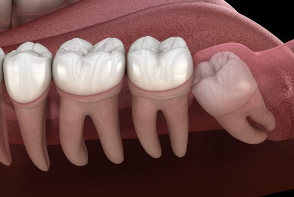
Develops around the crown of an unerupted tooth, most often wisdom teeth. Forms from tissue that normally surrounds a developing tooth. Requires surgical removal and usually extraction of the associated tooth.
Keratocyst
An aggressive cyst that grows rapidly and has high recurrence rates. Often affects the lower jaw near wisdom teeth. Requires careful surgical removal and close long-term follow-up to monitor for recurrence.
Gingival Cyst
Small cyst in the gum tissue, appearing as a tiny white or bluish bump. Common in infants but can occur in adults. Usually self-resolving in infants; surgical removal in adults if bothersome.
Nasopalatine Duct Cyst
The most common non-dental cyst in the mouth. Develops in the front of the roof of the mouth from remnants of embryonic tissue. Surgically removed if causing symptoms or actively growing.
Ranula
A mucocele that specifically forms on the floor of the mouth, often appearing as a large, translucent swelling under the tongue. Results from blocked salivary glands. Requires surgical removal of the cyst and associated gland.
Why Accurate Diagnosis Matters
- Different cysts require different treatments: Some need simple removal, others more extensive surgery
- Rule out serious conditions: Rare lesions can mimic benign cysts but require entirely different management
- Prevent recurrence: Correct identification ensures complete removal and reduces the chance of the cyst returning
- Plan appropriately: Knowing the type determines whether associated teeth need to be extracted alongside cyst removal
Signs & Symptoms
Symptoms vary depending on the type, size, and location of the cyst or mucocele. Many are completely asymptomatic in early stages, which is why regular dental checkups and X-rays are so important for early detection.
⚠️ See a Dentist If You Notice Any of These
- Visible bump or swelling in your mouth that persists more than 2 weeks
- Pain or pressure in the jaw or teeth
- Swelling that grows gradually over time
- Tooth displacement or mobility: teeth shifting or becoming loose
- Facial asymmetry or noticeable swelling of the face or jaw
- Difficulty eating, speaking, or swallowing
- Drainage or pus from the gums
- A mucocele that keeps coming back after rupturing
Oral Cyst Symptoms
What to Watch For
- Asymptomatic early on: Small cysts often cause no symptoms and are found incidentally on X-rays
- Swelling: Visible or palpable lump in the gum, jaw, or face as the cyst grows
- Pain or pressure: Discomfort in the affected area, especially if the cyst becomes infected
- Tooth movement: Teeth near the cyst may shift as it expands into surrounding bone
- Tooth discoloration: Affected tooth may darken if the nerve is dead
- Sensitivity: Temperature sensitivity or pain when chewing
Mucocele Symptoms
What to Watch For
- Visible bump: Soft, round, bluish or clear bump on the lip, cheek, or tongue
- Usually painless: Most mucoceles do not hurt unless irritated or infected
- Size fluctuation: May rupture, releasing clear fluid, then refill and grow again
- Functional interference: Large mucoceles can make eating, speaking, or wearing dentures difficult
- Chronic irritation: Repeatedly biting the same spot can trigger recurring mucoceles
How Are They Diagnosed?
Proper diagnosis ensures appropriate treatment and rules out more serious conditions. Our comprehensive diagnostic process at Best Dental covers all bases.
Clinical Examination
Visual and tactile inspection of the lesion. We assess size, location, consistency, color, and whether it is tender. Nearby teeth are also checked for signs of infection or trauma that may have triggered the cyst.
Dental X-Rays or CBCT Scan
Panoramic X-rays or cone beam CT scans reveal cysts within the jawbone, showing their size, boundaries, and proximity to teeth, nerves, and other structures. Mucoceles in soft tissue typically will not appear on X-rays.
Vitality Testing
For cysts associated with specific teeth, we test whether the tooth is alive or necrotic. This determines whether root canal therapy or extraction is needed alongside cyst removal.
Biopsy (During Removal)
Tissue removed during surgery is sent to a pathology lab for microscopic examination. This confirms the diagnosis and definitively rules out malignancy. Results typically return within one to two weeks.
Conditions We Differentiate From
- Abscesses: Pus-filled infections that are usually painful and require antibiotics plus drainage
- Fibromas: Firm, fibrous tissue growths that feel different on palpation
- Salivary gland tumors: Can mimic mucoceles but require entirely different treatment
- Oral cancer: Particularly important to rule out for lesions that do not heal or change appearance over time
Treatment Options
Treatment depends on the type, size, location, and symptoms of the cyst or mucocele. Our goal is always to use the most conservative effective approach, but in the majority of cases surgical removal is the right call.
When Can We Watch and Wait?
Conservative Management May Be Appropriate For
- Very small, asymptomatic cysts: Closely monitored with regular X-rays to track any growth
- Tiny mucoceles (under 3mm): May resolve on their own within 2 to 4 weeks
- Gingival cysts in infants: Epstein's pearls typically disappear without intervention
Most oral cysts and persistent mucoceles do require surgical removal. Cysts continue growing and can damage teeth, bone, and nerves. Mucoceles frequently recur unless the damaged salivary gland is also removed. Biopsy is needed to confirm the diagnosis. Leaving cysts untreated always makes future removal more complex.
Treatment by Cyst Type
Treatment Approaches at Best Dental
What About Marsupialization?
For very large cysts, a two-stage approach may be used. Stage one creates an opening to allow the cyst to drain and shrink over several months, decompressing it and stimulating bone regeneration. Stage two completes the surgical removal once the cyst has reduced significantly in size. This approach protects nearby teeth and nerves while achieving better long-term healing.
The Removal Procedure: What to Expect
Understanding what happens step by step reduces anxiety and ensures you are fully prepared. At Best Dental, your comfort is the priority throughout.
Before Surgery: Pre-Op Instructions
- Medical history: Inform us of all medications, allergies, and health conditions before your appointment
- Fasting: No food or drink for 6 to 8 hours if sedation is being used. Not required for local anesthesia only.
- Transportation: Arrange a driver if you are receiving sedation. You cannot drive yourself home.
- Medications: Continue regular medications unless specifically told otherwise
- Antibiotics: May be prescribed beforehand if the cyst is infected
Anesthesia
Local anesthesia is administered to ensure you feel no pain during the procedure, only pressure. For anxious patients, oral sedation or IV sedation ($500 flat per session) is available to keep you calm and relaxed throughout. General anesthesia is rarely needed except for very extensive cases.
Incision and Access
An incision is made in the gum or oral tissue to access the cyst or mucocele. For cysts in the jawbone, a small amount of overlying bone may be removed to fully expose the lesion.
Cyst or Mucocele Removal
For mucoceles, the cyst and the damaged salivary gland are carefully excised together to prevent recurrence. For oral cysts, the entire cyst lining is removed and the cavity curetted to ensure no tissue remains. Large bone cavities may be filled with graft material to support healing.
Associated Tooth Treatment (If Required)
If a tooth is involved in the cyst, it may receive root canal therapy to save it, or be extracted if it is non-restorable. We always pursue tooth preservation when possible and discuss all options with you before proceeding.
Closure and Post-Op Instructions
The incision is closed with dissolvable sutures and gauze is placed to control initial bleeding. Written post-operative instructions are provided before you leave. Most patients comfortable enough to drive home (without sedation) and can resume light activity the following day.
Recovery & Aftercare
Recovery from oral cyst or mucocele removal typically takes one to two weeks, though most patients feel significantly better within three to five days. The key is following post-operative instructions carefully in the first 48 hours.
⚠️ Critical First 24 to 48 Hours
- Bleeding: Some oozing is normal. Bite on gauze firmly for 30 to 60 minutes. If bleeding continues, a damp tea bag works well as tannic acid aids clotting.
- Swelling: Peaks at 48 to 72 hours. Use ice packs for the first 24 hours, then switch to warm compresses.
- Pain: Take prescribed pain medication before the numbness wears off. Staying ahead of pain is much easier than catching up to it.
- Diet: Soft, cool foods only. Avoid hot liquids, hard or crunchy foods, and drinking through straws.
Days 3 to 7
What to Expect
- Swelling decreases: Should be noticeably reduced by day 5
- Stitches dissolve: Dissolvable sutures fall out on their own. Do not pull them.
- Resume normal diet: Gradually reintroduce regular foods as comfort allows
- Oral hygiene: Resume gentle brushing, avoiding the surgical site initially
- Saltwater rinses: Begin gentle rinses 24 hours after surgery: half a teaspoon of salt in 8 oz warm water
Weeks 2 to 4 and Beyond
Longer-Term Healing
- Soft tissue heals: Surgical site fully closes in 2 to 3 weeks
- Bone regeneration: For cysts in the jaw, bone healing takes 3 to 6 months and is monitored with follow-up X-rays
- Follow-up appointment: We review your biopsy results, assess healing, and confirm no signs of recurrence
- Return to full activity: Most patients resume exercise and normal routines within 1 to 2 weeks
⚠️ Call Us Immediately If You Experience
- Excessive bleeding that does not stop with sustained pressure
- Severe pain not controlled by prescribed medication
- Fever over 101 degrees F or chills
- Swelling that worsens after day 3 instead of improving
- Pus or foul discharge from the surgical site
- Difficulty breathing or swallowing: go to an emergency room immediately
- Numbness persisting well beyond the expected anesthesia duration
Frequently Asked Questions
No. The vast majority are completely benign. However, biopsy during removal is standard practice to confirm the diagnosis and definitively rule out malignancy. Rarely, some cysts can harbor concerning features, which is exactly why removal and pathology examination matters even when a lesion appears benign on the surface.
Recurrence rates are low when the cyst is completely removed with its full lining intact. Mucoceles recur in about 10 to 15 percent of cases if the damaged salivary gland is not fully excised alongside the cyst. Certain aggressive types like keratocysts have higher recurrence rates and require closer long-term follow-up.
The procedure itself is painless. Local anesthesia ensures you feel only pressure, not sharp pain. Afterward, expect mild to moderate discomfort for 3 to 5 days, which is well-controlled with over-the-counter or prescribed pain medication. Most patients describe the recovery as less painful than a tooth extraction.
No. While mucoceles may rupture on their own, they almost always refill within hours or days because the underlying damaged salivary gland continues producing saliva. Attempting to pop it yourself risks introducing infection and does not address the root cause. Proper surgical removal of both the mucocele and the damaged gland is needed for permanent resolution.
Most patients take 1 to 2 days off for simpler procedures like mucocele removal or small cysts. Larger cyst removals may require 3 to 5 days of rest. Most patients can return to desk work or school within 2 to 3 days, avoiding strenuous physical activity for about a week.
Tooth preservation is always our primary goal. For cysts not involving specific teeth, surrounding teeth are typically unaffected. For cysts associated with a particular tooth, that tooth may need root canal therapy or extraction, but adjacent teeth generally remain intact. We walk through all options with you before any treatment begins.
Yes. Removal of oral cysts and mucoceles is typically covered by dental insurance as a medically necessary procedure, not a cosmetic one. Coverage usually ranges from 50 to 80 percent after your deductible. Best Dental is in-network for all 8 major PPO carriers. Our team verifies your benefits before treatment and provides a written cost estimate before you commit.
Any bump, swelling, or lesion that persists for more than 2 weeks should be professionally evaluated. Do not wait to see if it resolves on its own. Early diagnosis leads to simpler treatment, less invasive surgery, and better outcomes. Call us at (281) 215-3065 to schedule an evaluation.
Key Takeaways: Oral Cyst & Mucocele Removal
Expert Treatment for Oral Cysts & Mucoceles
Do not ignore bumps, swelling, or lesions in your mouth. At Best Dental in Richmond, TX, we provide comprehensive evaluation and expert surgical removal, conveniently located for Houston, Sugar Land, Katy, and Fort Bend County. Mucocele removal: $500 flat. Early treatment prevents complications and ensures the best outcomes.